Next: Research projects No:34-44
Up: Research
Previous: Research projects No: 12-22
- Wood fibre morphology
Mattias Moëll, Gunilla Borgefors
Funding: Wood and Wood Fiber graduate school; SLU S-faculty
Period: 9509-0203
Partners:
Lloyd L. Donaldson, Forest Research New Zealand Ltd., Rotorua, New Zealand;
Minoru Fujita, Graduate School of Agriculture, Kyoto University, Japan
Abstract:
The morphology of wood fibres is of great importance to the mechanical
properties of pulp and paper. For the forest industry to be able to produce
new products, renew processes, and to maximise the use of the wood fibre
potential, more knowledge of the fibre morphology is needed. The project
concentrated on analysis of fibre cross-sections in confocal microscopy
images of transverse sections of wood. The aim was to measure as many
parameters as possible, such as: cell wall width, radial/tangential lumen
width, fraction of cell wall area, and degree of compression wood. A fully
automatic image analysis method was developed, where each individual fibre
was measured, and the measures are averaged along the wood section. The
same method can be used for different wood species by changing a few
parameters. In December 2001, Moëll successfully defended his PhD thesis.
- Analysis of AFM images of wood fibres using image analysis
algorithms
Carolina Wählby
Funding: UU TN-faculty
Period: 0101-
Partners:
Jesper Fahlén, STFI, Swedish Pulp and Paper Research Institute, Stockholm
Abstract:
Understanding the arrangement of wood polymers within the fibre wall is
important for understanding the mechanical properties of the fibres
themselves. Due to their high load bearing ability, the arrangement of
cellulose fibrils within the cell wall are of special interest. In this
work AFM-Atomic Force Microscopy-in combination with image analysis
algorithms originally developed for cell segmentation has been used to
obtain more information about the arrangement of cellulose aggregates
(fibrils) in the secondary cell wall layer of spruce wood. The effects of
chemical processing on the arrangement of these cellulose aggregates were
also studied. Enlargement of cellulose aggregates was found in the initial
phase of the kraft cook. This increase in cellulose aggregate dimensions
depended mostly on temperature for treatment temperatures above
140°C, regardless of the amount of alkali present. Although
hemicelluloses are lost to various degrees under alkaline conditions, the
increase in cellulose aggregate size was mainly related to thermally
induced rearrangement of the cellulose molecules. The mean side length of
cellulose aggregates was found to be around 18 nm in unprocessed wood and
23 nm in processed wood. The cellulose aggregates were assumed to be square
shaped in cross section in both cases.
- 3D tracking of fibres in paper
Mattias Aronsson, Stina Svensson, Gunilla Borgefors
Funding: Swedish Foundation for Strategic Research, VISIT
programme, SLU S-fak
Period: 9710-
Partners: Björn Kruse, Arash Fayyazi, Dept. of Science and
Engineering, Linköping University, Campus Norrköping; Örjan Sävborg, Olle
Henningsson, StoraEnso Research, Falun; Per Nygård, Cristine Antoine, Rune
Holmstad, Norwegian Pulp and Paper Research Institute (PFI), Trondheim, Norway
Abstract:
Using image analysis on paper samples can increase the understanding of the
individual fibres build up the paper and what effects different types of
fibre networks have on paper properties. This network of fibres is a very
complicated structure and creating images of it is a challenging problem.
It is essential to use 3D volume images, since 2D images cannot capture
enough information of the fibre network. Fibres are thin, so the resolution
must be in the micrometer range, to enable accurate measurements. Our main
concern is developing the necessary imaging and analysis tools to enable a
practical process method for creating volume images of paper samples, and
then use these images to measure various properties of the fibre network.
The main data set used is a volume image created from a series of 2D
scanning electron microscopy (SEM) images captured at StoraEnso Research,
Falun. We have now created a rather large digital volume of a paper sample
from the original data set, see cover page, for a visualisation of a small
part of the paper volume.
As fibre curvature effects the mechanical properties of paper, we are
developing methods to estimate the curvature. The theoretical work, i.e.,
curvature estimations for voxels in a discrete curve, is described in
Project 41. Initial results, where the curvature estimator
was used in the analysis of the fibres, were presented at the SSAB
symposium in Lund. Continuing our efforts to develop three dimensional
measurements, we added a method for twist and aspect ratio estimation,
which was presented at ICPR in Québec, Canada. A previous fibre detection
algorithm assumed a quadratic grid. This was generalised and the new method
developed was presented at ICIP in Rochester, NY, USA. A number of
different types of fibre and fibre network measurements have also been
developed. Many of the measurements are based on using distance
transformations (see Project 37) of the fibres, fibre walls,
fibre lumens, and the paper pores. An article where we describe many
distance based measures was accepted for publication in a special issue of
IEEE Transactions on Systems, Man and Cybernetics, and will be published
during 2003.
For Aronsson, preparations for the PhD thesis dominated this year's work.
This included finalising and description of the progress and the insights
gathered during the project in a structured manner. On 12 December 2002,
Aronsson successfully defended his thesis entitled ``On 3D Fibre
Measurements of Digitized Paper - from Microscopy to Fibre Network''. See
Section 4.3.
- Analysing the structure of paper sheet through confocal microscope
images
Ingela Nyström
Funding: UU TN-faculty
Period: 0205-
Partners: Catherine Östlund, Swedish Pulp and Paper Research
Institute (STFI), Stockholm
Abstract:
It is of interest to study the structure of paper surface under different
pressure to measure how the pores change. One possible way is to produce
confocal microscope images of paper sheet and analyse these. The original
three-dimensional (3D) images consist of, in principle, cubic voxels which
can be transformed to two-dimensional (2D) grey-level images, where the
grey-levels correspond to the depth at which the first paper fibre is
visible, i.e., how deep the pores penetrate into the paper.
Initially, we have analysed three such 2D grey-level images, where the
pressure has been low, medium, and high, respectively. The pressure may be
unevenly distributed, a problem overcome by computing a grey-level convex
hull (see Project 35).
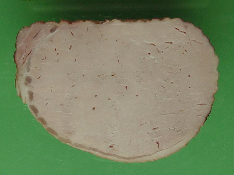
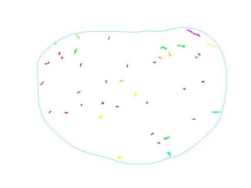
- Image analysis methods for food quality measurements
Lucia Ballerini, Gunilla Borgefors
Funding: Foundation for Strategic Environmental Research (MISTRA),
FOOD 21 programme
Period: 9908-0209
Partners: Dept. of Food Science, SLU, Uppsala; Wallenberg
Laboratory, Sahlgrenska University Hospital, Göteborg
Abstract:
FOOD 21 is a broad scientific project, aimed to develop sustainable food
production methods. Image processing methods have been successfully applied
to meat images in order to determine the percentage and the distribution of
fat and various defects. We have been working with camera photographs and
Magnetic Resonance images. Segmentation algorithms have been optimised for
these kinds of images, in order to classify different substances as muscle,
fat and connective tissue. Moreover, we developed methods to measure
homogeneity of fat distribution. Indeed, fat distribution is an important
criterion for meat quality evaluation and its expected palatability. This
method is simple and accurate and gives a description of feature
distribution and a measure of homogeneity, depending on both size and
spatial organisation of features, without requiring any individual measures
of them. Similar segmentation techniques and distribution measurements have
also been developed for defects in processed meat, such as pores, holes,
and cracks. See Figure 10. Different meat processing methods have
been evaluated using these results. This year the project has produced one
article in ``IEEE Transactions on Nuclear Science'' and three conference
papers.
Figure 10:
Digital camera image of pig meat and extracted holes (holes are coloured
based on their size).
- New techniques for information extraction by using new neuro-fuzzy systems
Hamed Hamid Muhammed
Funding: UU TN-faculty, Swedish National Space Board
Period: 0201-
Abstract:
New neuro-fuzzy systems, which imitate the functionality of the biological
visual system, were developed in this work based on the new so-called
Weighted Neural Networks (WNN). The basic idea of WNNs, is to modify
well-known Artificial Neural Networks (ANN) by additional mechanisms to be
able to capture/calculate and store as much useful information as possible
about the input data set at hand. So far, two main types of WNNs can be
recognised: incremental and fixed (or grid-partitioned) depending on the
original ANN algorithm used as a start point. It seems possible to make a
WNN version for every existing ANN. The WNN algorithm (incremental or
grid-partitioned) produces a net of nodes connected by edges. Additional
weights, which are proportional to the local densities in the input space,
are associated with the resulting nodes and edges to store useful
information about the topological relations in the given input data set. A
fuzziness factor, proportional to the connectedness of the net, is
introduced in the system. The basic idea is based on the famous Hebb's
postulate which states that the connection between two winning neurones
gets stronger. The result is a weighted connected net, consisting of
weighted nodes connected by weighted edges, which reflects and preserves
the topology of the input data set, and in addition to that, it acts as a
fuzzy representation of the input data set. Two main types of WNNs have
been recognised, so far:
- Weighted Fixed Neural Networks (WFNN):
The basic idea here is to distribute a number of zero-weighted nodes, as an
equidistant initial grid in input space where the input data set is found.
Then, weights are assigned to these nodes, where a relatively high
node-weight corresponds to a relatively high density in a neighbourhood
around the node in input space. In addition to that, the algorithm connects
neighbouring nodes with weighted connections or edges, where an edge-weight
is also proportional to the density of input data in the region between the
connected nodes (or in a neighbourhood around the edge). A fuzziness factor
is introduced here as mentioned above. The work has resulted in a refereed
journal paper presenting the WFNN algorithm.
- Weighted Incremental Neural Networks (WINN):
The WINN is an incremental self-organising model with no pre-defined
structure, and therefore no restrictions on the dimensionality of the input
data set, which can have different dimensions in different regions of input
space. The model is built by successive addition, adaptation, and sometimes
deletion of elements (i.e., nodes and edges), according to suitable
strategies, until a stopping criterion is met. Here also, a weighted
connected net, which preserves the topology of the input data set, is
produced. The algorithm begins with only two nodes connected by an edge,
then new nodes and edges are generated and the old ones are updated (and
sometimes deleted) while the learning process proceeds until a certain
stopping criterion is met. Here also, a fuzziness factor is introduced here
as mentioned above when talking generally about WNNs.
- Genetic snakes
Lucia Ballerini
Period: 0107-0209
Abstract:
Genetic Snakes are active contour models, also known as snakes, with an
energy minimisation procedure based on Genetic Algorithms. Genetic Snakes
have been proposed to overcome some limits of the classical snakes and
successfully applied to medical, radar and meat images. During 2002, they
have been extended by using a new form of the image energy which considers
texture features. They have been applied to segment liver in CT images,
which was presented at EMBEC in Vienna in December 2002). The model has
also been extended by adding an elastic force that couples multiple
contours together and create what we call ``multiple genetic snakes''. This
model has been used to segment bones in hand radiograms, which will be
presented at SPIE Medical Imaging in San Diego in February 2003. A further
extension, currently under study, is the evolution of the weights and the
functionals that control the snake behaviour, i.e., the internal energy
determined by the elasticity and rigidity of the snake, and the image
energy representing the characteristics of the image (intensity, gradient,
etc.).
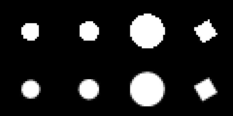
- Modelling of natural objects
Felix Wehrmann, Ewert Bengtsson
Funding: UU TN-faculty
Period: 9912-
Abstract:
This project started under the scope of the general idea of model-based
segmentation. A large number of images, especially from the medical sector,
lack a proper description of the objects the image analyst is interested
in. Often, this leads to poor results of automated segmentation procedures,
if any. Incorporating information about the shape of an object is one
possible completion of an object description. However, models that provide
shape descriptions usually lack the power to compensate for the variation
nature supplies us with. As an example, we could ask ourselves, which
features make us easily recognise and localise a brain in a medical
3D-image, a task which has automated solutions only in specific cases.
With the intention to compensate for natural variation, we applied a number
of common concepts to the problem. In particular, orthogonal transforms,
such as PCA and ICA, have been inspected in an attempt to derive the
characteristic correlations between similar shapes. Moreover, the
applicability of Markov random fields as a stochastic modelling concept was
analysed.
It turned out that a general model should not be dependent on landmarks as
required for the previous transformations. Since variations in
landmark-less shape data appear as non-linear manifolds, a neural network
was designed to acquire the particularities of the data. After training on
examples, the network provides a non-linear representation of shape by
means of its modes of variation. So far the representation has been tested
on simple shapes and variations showing promising results, see
Figure 11.
Further characteristics of non-linear models should be examined in the
future, as for example the possibilities of shape decomposition.
Figure 11:
A neural network learns the variation of a clover-like shape. The shapes
are produced by the network after learning the essentials from examples.
Stacked together, they leave the impression of a vase.
- Global shape description in 2D and 3D by polynomial expansion
Ola Weistrand, Gunilla Borgefors
Funding: TFR; UU TN-faculty
Period: 9701-
Partners: Christer Kiselman, Dept. of Mathematics, UU; Örjan
Smedby, Dept. of Medicine and Care, Linköping University Hospital
Abstract:
Shape description derived from volume images is usually local, e.g., finite
elements, surface facets, and spline functions. This can be a severe
limitation on usefulness, as comparison between different shapes becomes
very difficult. In 2D, Fourier descriptors is a successful and often used
global descriptor with adaptable accuracy. This concept cannot be
immediately generalised to 3D because it relies heavily on the existence of
an ordering of the boundary pixels. The aim of this project is to overcome
this problem and develop methods for global shape description in 3D. At the
moment we study a limited class of objects, those that are homotopic to the
sphere. By recursively morphing a sphere to the surface of the object, a
parametrisation of the object is obtained from a parametrisation of the
sphere. The method is computationally attractive. Using the object
parametrisation we can approximate coordinate functions using a linear
combination of spherical harmonics, that is a complete orthonormal set of
functions on the unit sphere. By aligning the coordinate system along the
principal axis of the object we hope to obtain approximately invariant
coefficient for objects differing only by a combination of translations and
rotations
- Accurate and precise size estimators for digitised 2D and 3D
objects using local computations
Joakim Lindblad, Ingela Nyström
Funding: UU TN-faculty
Period: 0012-
Partners: Jayaram K. Udupa, MIPG, Dept. of Radiology, University of Pennsylvania, Philadelphia, USA
Abstract:
Information is irrevocably lost in the process of digitising a continuous
object of the real world to fit the digital world of the computer.
Therefore, feature measurements of digitised objects can be no more than
estimates. Good estimators are those that approach the corresponding
feature value of the continuous original object. The possibility to use
only local computations is a desirable property in computerised image
analysis, both to keep the complexity level at a minimum, and to enable for
parallelism in various ways. This project aims at finding good local
estimators for size related measures of digitised objects, i.e., perimeter
and area of 2D objects, and surface area and volume of 3D objects.
Statistical validation of the estimators have been performed on large
numbers of computer generated digitised objects. The breakdown behaviour at
very low resolution, as well as the asymptotic behaviour at high resolution
have been studied.
A surface area estimator with improved precision and accuracy obtained by
optimising the area contribution locally, was presented at the DGCI 2002
conference in Bordeaux.
The work on enclosed volume of triangulated surfaces, which can be computed
efficiently, in the same elegant way similarly to digital surface
integration, simultaneously with surface area computation, was presented at
SPIE Medical Imaging 2002.
This projects relates to Project 33 on shape of fuzzy sets.
- Fuzzy shape analysis in 2D and 3D
Nataša Sladoje (Matic), Ingela Nyström, Gunilla Borgefors
Funding: SLU S-faculty, UU TN-faculty
Period: 0109-
Partners: Punam K. Saha, MIPG, Dept. of Radiology, University of Pennsylvania, Philadelphia, USA
Abstract:
Fuzzy segmentation methods, that have been developed in order to reduce the
negative effects of the unavoidable loss of data in the digitisation
process, initialise the interest for new shape analysis methods, handling
grey-level images. Very little has been published to date on shape analysis
of fuzzy segmentations. We have performed initial studies on perimeter and
area of 2D fuzzy subsets, where the focus have been on objects with fuzzy
border. We assume that in the segmentation process most pixels easily can
be classified either as object or background, but for pixels located in the
vicinity of the boundary of the digitised object it is hard to make such a
discrimination. One way to treat these pixels is to determine the extent of
their membership to the object. The membership value of a pixel can be
defined as the fraction of its area that belongs to the original object,
see Figure 12. We have implemented a method where we propose
perimeter and area estimators adjusted to the discrete case. The estimates
are computed for a large collection of fuzzy segmented digitised objects.
We have concluded that our method highly improves both accuracy and
precision of the results obtained from crisp (hard) segmentation, especially
in the case of low resolution images, i.e., small objects.
As the next step, we will investigate different ways to extend the main
binary shape analysis tools (e.g., distance transform, medial axis, notion
of convexity, and moments) to fuzzy segmented 2D and 3D images.
Figure 12:
Examples of digitised objects with crisp (top)
and fuzzy border (bottom).
Next: Research projects No: 34-44
Up: Research
Previous: Research projects No:12-22