Diffraction Artifact Reduction in µCT Imaging
Erik Wernersson, Cris Luengo, Anders Brun, Gunilla Borgefors
Partners: Jan Van den Bulcke, Dept. of Forest and Water Management, Ghent University, Belgium; Matthieu Boone, Dept. of Physics and Astronomy, Ghent University, Belgium
Funding: S-faculty, SLU
Period: 1009-1412
Abstract: When imaging wood based materials, diffraction causes artefacts especially around sharp
edges. While sometimes useful, and the only measurable properties of the imaged objects, they
might as well be a nuisance which hinders proper analysis of the absorption coefficient. In this
project, different ways to reduce such artefacts are investigated, especially in already reconstructed
images. Compare to previous approaches, this is much faster and does not require that the original
projection images are stored.
This year we presented a paper at SSBA that showed how to tune the parameters of the method that we published in the Journal of the Optical Society of America A (2013). Erik Wernersson defended his PhD thesis closely related to this project in December 2014.
Image Analysis of the Internal Structure of Paper and Wood Fibre Based Composite Materials in 3D images
Erik Wernersson, Anders Brun, Cris Luengo, Gunilla Borgefors
Partners: Gary Chinga, Norwegian Pulp and Fibre Research Institute, Trondheim, Norway;
Catherine Östlund, Innventia, Stockholm; Thomas Joffre, Dept. of Engineering Sciences, Applied
Mechanics, UU; Arttu Miettinen, Dept. of Physics, University of Jyväskylä (UJ), Finland; Joakim Lindblad, University of Novi Sad, Serbia; Svetlana Borodulina, Dept. of Solid Mechanics and BiMaC Innovation Center, KTH
Funding: S-faculty, SLU; WoodWisdom-Net
Period: 0406-1412
Abstract: The internal structure of paper is important because many of its properties correspond
directly to the properties of single fibres and their interaction in the fibre network. How single
fibres in paper bond and how this affects paper quality is not fully understood, since most structure
analysis of paper has been performed in cross-sectional, two-dimensional (2D) images whereas
paper is a complex, three-dimensional (3D) structure.
Another application for wood fibres that has recently gained interest is wood polymer
composite materials. The properties of these materials do not only depend on the structure of the fibre
network, but also on the interaction between the fibres and the polymer matrix surrounding the
fibres.
Advances in imaging technology have made it possible to acquire 3D images of paper and wood polymer composite materials. In this project, image analysis methods for characterising the 3D material structure in such images are developed. The detailed knowledge of the material structure attainable with these methods is useful for improving material properties and for developing new materials.
The project objective is to achieve a complete segmentation of individual fibres and pores in volume images of the material. Given such a segmentation, any desired measurement of the internal structure is available. A sample segmentation result is shown in Figure 18. Measurements on individual fibres and the structural arrangement of fibres can then be related to macroscopic material properties.
In this project, different volume images of paper and composite materials are available: one volume created from a series of 2D scanning electron microscopy (SEM) images at StoraEnso, Falun; and X-ray microtomography volume images of paper and composite samples imaged at the European Synchrotron Radiation Facility (ESRF) in Grenoble, France, at the Paul Scherrer Institut (PSI) in Villigen, Switzerland and also from tabletop scanners at University of Jyväskylä, Finland, at Applied Mechanics, Uppsala University, and at Innventia, Stockholm.
This year we published papers in the Nordic Pulp & Paper Research Journal, Cellulose, and the Mechanics of Materials. Erik Wernersson defended his PhD thesis closely related to this project in December 2014.
|
|
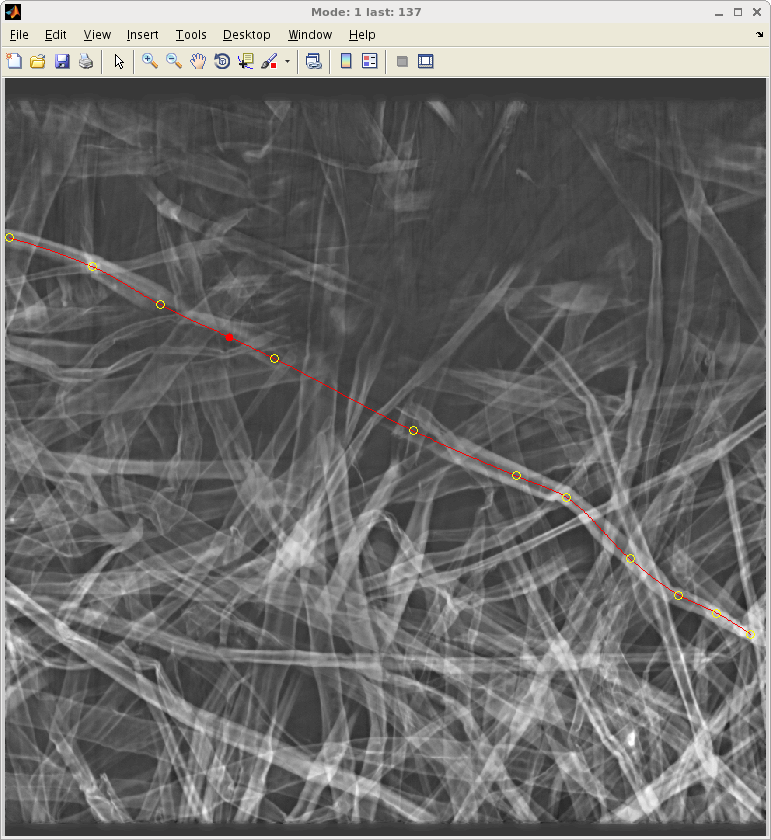
Ring Width and Density Profiling with Helical CT
Erik Wernersson, Cris Luengo, Anders Brun, Gunilla Borgefors
Partners: Jan Van den Bulcke, Dept. of Forest and Water Management, Ghent University, Belgium
Funding: S-faculty, SLU
Period: 1201-1412
Abstract: Dendrochronology relies on accurate measurements of annual ring widths. The most
common method is to use a flatbed scanner to acquire high resolution images of polished wood
surfaces. In this project we investigate potential gains using a helical X-ray device which produces
volume images. Direct advantages include non destructive and simplified sample preparation procedures
as well as compensation for the orientation of the inner structure which can not be seen
with ordinary flatbed scans. It is also possible to find density profiles using the same images.
One article was published in Dendrochronologia. Erik Wernersson defended his PhD thesis closely related to this project in December 2014.
Light Scattering in Paper
Erik Wernersson, Cris Luengo
Partners: Tomas Linder and Torbjörn Löfqvist, Luleå University of Technology, Luleå
Funding: S-faculty, SLU
Period: 1212-1412
Abstract: Fibre orientation is an important structural property in paper and other fibrous materials.
In this study we explore the relation between light scattering and in-plane fibre orientation
in paper sheets. Light diffusion from a focused light source is simulated using a
Monte Carlo technique where parameters describing the paper micro-structure were determined
from 3D x-ray computed tomography images. Measurements and simulations on both spatially
resolved reflectance and transmittance light scattering patterns show an elliptical shape
where the main axis is aligned towards the fibre orientation.
Good qualitative agreement was found at low intensities and the results indicate that fibre orientation in thin fibre-based materials can be determined using spatially resolved reflectance or transmittance. Published in Optics Express. Erik Wernersson defended his PhD thesis closely related to this project in December 2014.
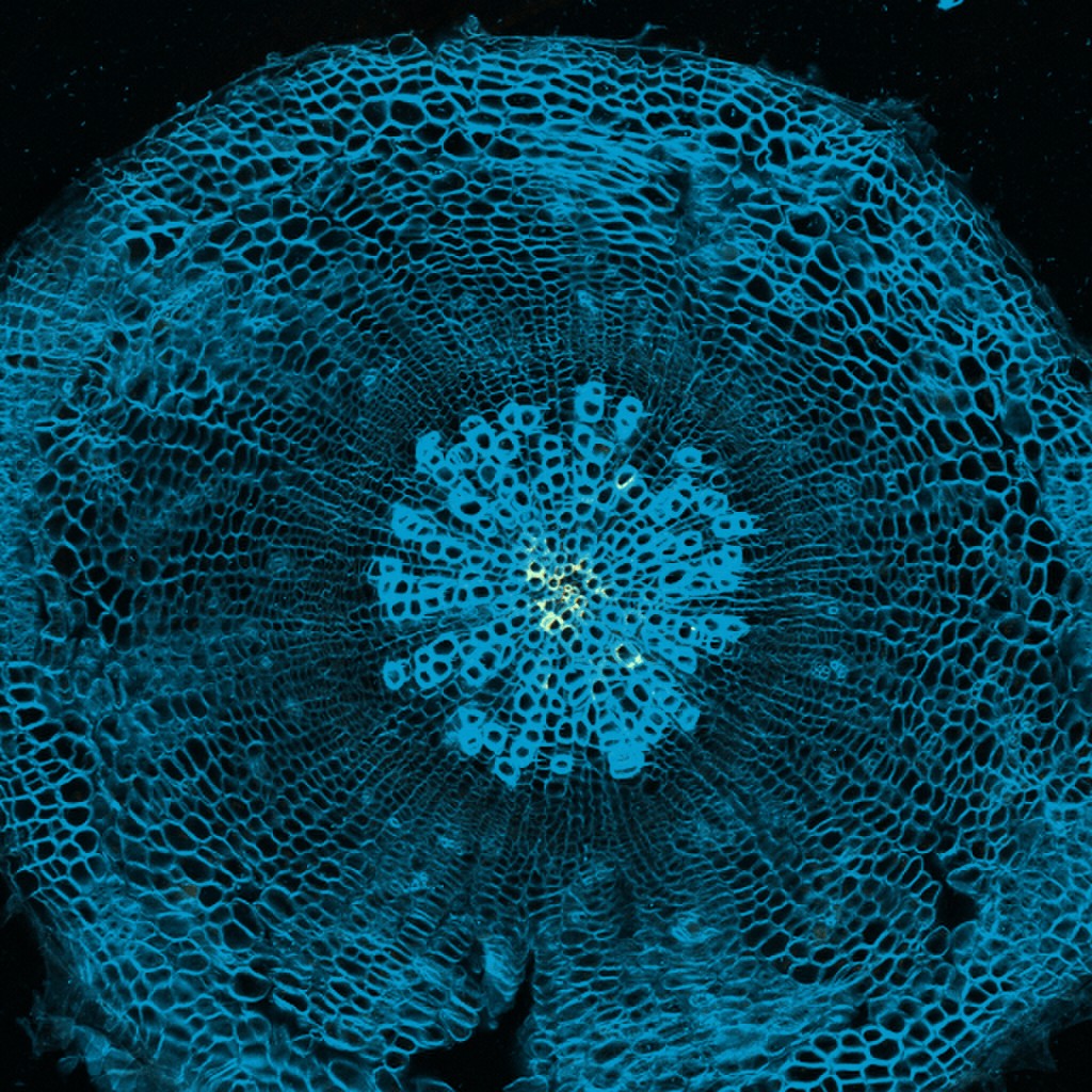
Large-scale quantification of gene expression in Arabidopsis
Azadeh Fakhrzadeh, Cris Luengo
Partners: Urs Fischer, Hardy Hall, Umeå Plant Science Centre, SLU
Funding: S-faculty, SLU
Period: 1402-
Abstract: Arabidopsis is the most important plant model organism. For animal model organisms such as Drosophila melanogaster (fruitfly), C. elegans
(roundworm) and Danio rerio (zebrafish), efforts have been made to map gene
expression on a per-cell or sub-cell resolution. In this project, we develop tools to create the first such map for a plant species. We prepare thin sections of the root, hypocotyl and stem of the plant at various stages between sprouting and maturity. These sections are fluorescently stained such that the cell
walls can be visualized in the confocal microscope. Each section also receives a FISH (fluorescent in situ hybridization) stain for a particular protein.
Sections are then imaged at a magnification that allows most of the section to fit in the field of view. This yields several thousand cells in each image. Next, we use a fully automatic segmentation and quantification pipeline that allows measurement of relative amount and quality of the stained protein in each
subcellular area (wall and lumen are separated, and each divided into four regions: inner, outer and two lateral). Cells are automatically classified into the various cell types, which allows statistics of expression over each of the cell types, for example. We currently have imaged several thousand sections, from both wildtype and mutant samples, stained for hundreds of different genes (Figure 19).
|
|