
|
Bone anchored implants, both for oral- and orthopaedic purposes, are important and frequently used as spare parts in the body. An important step in the understanding process of the mechanisms of of bone-implant integration is evaluation of regenerated bone tissue in the proximity of implants.
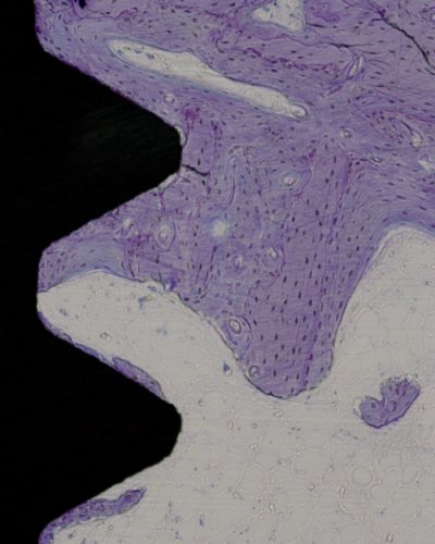
Until recently, implant integration was only evaluated from 2D microscopic
images. Reliable analysis of bone-implant integration requires 3D imaging as the whole sample should be included in the analysis.
In this project we present methods for visualization of SRμCT-scanned 3D volumes of screw-shaped bone implant samples. Two presented methods are thread fly-through and 2D unfolding.
|

