Hamid Sarve - 2D unfoldings
2D Unfolding of Bone-implant Samples
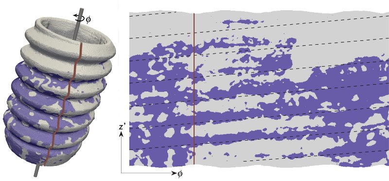
The 2D unfolding is performed by a radial projection of the relevant feature
information onto the implant surface, followed by an angular sampling.
Figure below show:
Top: rendered surface of the implant with bone-implant contact regions
superimposed and the unfolded surface IBC. Black dashed lines show the
approximate location of the peaks of the threads. The vertical line indicates the corresponding
angles in the two images.
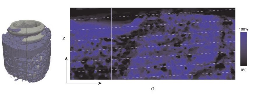
Bottom: Rendered surface of the implant with bone tissue volume in
the region of interest superimposed and the unfolded surface. White dashed
lines show the peaks of the threads. The vertical line indicates the corresponding angles
in the two images.