next up previous contents
Next: Analysis of microscopic biomedical Up: Research Previous: Research   Contents

Forestry related applications

  1. Diffraction Artifact Reduction in µCT Imaging
    Erik Wernersson, Cris Luengo, Anders Brun, Gunilla Borgefors
    Partners: Jan Van den Bulcke, Dept. of Forest and Water Management, Ghent University, Belgium; Matthieu Boone, Dept. of Physics and Astronomy, Ghent University, Belgium
    Funding: S-faculty, SLU
    Period: 1009 -
    Abstract: When imaging wood based materials, diffraction causes artifacts especially around sharp edges. While sometimes useful, and the only measurable properties of the imaged objects, they might as well be a nuisance which hinders proper analysis of the absorption coefficient. In this project, different ways to reduce such artifacts are investigated, especially in already reconstructed images. Compare to previous approaches, this is much faster and does not require that the original projection images are stored. For an example of the artifact, see Fig. 2.

    We have had one article published in Journal of the Optical Society of America during 2013. One of the main results is that it is at least as good to remove the diffraction artifacts after the reconstruction as before it.

    Figure 2: A slice from a volume image of a paper sample. (a) directly reconstructed, a mixed imaged with both phase and amplitude. (b) phase contribution removed to reveal the amplitude or absorption.
    Image org Image filt

  2. Image Analysis of the Internal Structure of Paper and Wood Fibre Based Composite Materials in 3D images
    Erik Wernersson, Anders Brun, Cris Luengo, Gunilla Borgefors
    Partners: Gary Chinga, Norwegian Pulp and Fibre Research Institute, Trondheim, Norway; Catherine Östlund, Innventia, Stockholm; Thomas Joffre, Dept. of Engineering Sciences, Applied Mechanics, UU; Arttu Miettinen, Dept. of Physics, University of Jyväskylä (UJ), Finland; Joakim Lindblad, University of Novi Sad, Serbia; Svetlana Borodulina, Department of Solid Mechanics and BiMaC Innovation Center, KTH
    Funding: S-faculty, SLU; WoodWisdom-Net
    Period: 0406-
    Abstract: The internal structure of paper is important because many of its properties correspond directly to the properties of single fibres and their interaction in the fibre network. How single fibres in paper bond and how this affects paper quality is not fully understood, since most structure analysis of paper has been performed in cross-sectional, two-dimensional (2D) images whereas paper is a complex, three-dimensional (3D) structure.

    Another application for wood fibres that has recently gained interest is wood polymer composite materials. The properties of these materials do not only depend on the structure of the fibre network, but also on the interaction between the fibres and the polymer matrix surrounding the fibres.

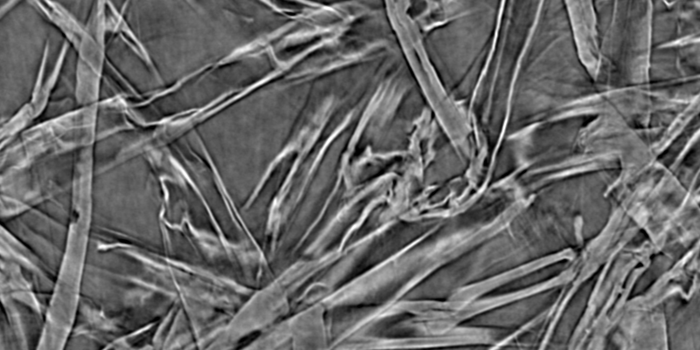
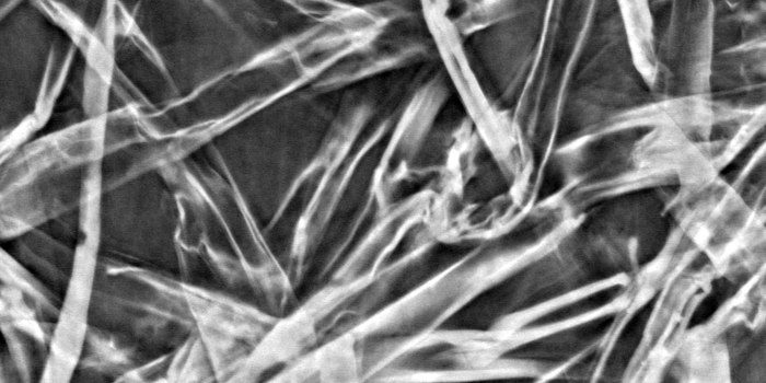
    Advances in imaging technology have made it possible to acquire 3D images of paper and wood polymer composite materials. In this project, image analysis methods for characterizing the 3D material structure in such images are developed. The detailed knowledge of the material structure attainable with these methods is useful for improving material properties and for developing new materials.

    The project objective is to achieve a complete segmentation of individual fibres and pores in volume images of the material. Given such a segmentation, any desired measurement of the internal structure is available. Measurements on individual fibres and the structural arrangement of fibres can then be related to macroscopic material properties.

    In this project, different volume images of paper and composite materials are available: one volume created from a series of 2D scanning electron microscopy (SEM) images at StoraEnso, Falun; and X-ray microtomography volume images of paper and composite samples imaged at the European Synchrotron Radiation Facility (ESRF) in Grenoble, France, at the Paul Scherrer Institut (PSI) in Villigen, Switzerland and also from tabletop scanners at University of Jyväskylä, Finland, UU, and Innventia, Stockholm.

  3. Generation of Synthetic µCT Volumes
    Erik Wernersson, Cris Luengo, Anders Brun, Catherine Östlund, Gunilla Borgefors
    Partners: Norwegian Pulp and Paper Research Institute (PFI), Trondheim, Norway; Innventia, Stockholm; Dept. of Engineering Sciences, Applied Mechanics, UU; Dept. of Physics, University of Jyväskylä (UJ), Finland; SINTEF Materials and Chemistry, Norway; Risø National Laboratory, Technical University of Denmark
    Funding: S-faculty, SLU; WoodWisdom-Net
    Period: 0901-
    Abstract: It is of great importance to evaluate the performance and stability of new methods. It is often hard to do so, when working with natural materials, since no true answer is available. With this project we aim to create highly realistic reference images that can be used to evaluate new and existing methods designed for characterisation of fibrous materials from µCT.

    Within the project, methods have been developed to generate and pack synthetic wood fibres as well as to simulate µCT acquisition systems with characteristic artifacts.

  4. Ring Width and Density Profiling with Helical CT
    Erik Wernersson, Cris Luengo, Anders Brun, Gunilla Borgefors
    Partners: Jan Van den Bulcke, Dept. of Forest and Water Management, Ghent University, Belgium
    Funding: S-faculty, SLU
    Period: 1201 -
    Abstract: Dendrochronology relies on accurate measurements of annual ring widths. The most common method is to use a flatbed scanner to acquire high resolution images of polished wood surfaces. In this project we investigate potential gains using a helical xray device which produces volume images. Direct advantages include non destructive and simplified sample preparation procedures as well as compensation for the orientation of the inner structure which can not be seen with ordinary flatbed scans. It is also possible to find density profiles using the same images. During 2013, one article was submitted to Dendrochronologia which will be published during 2014.


next up previous contents
Next: Analysis of microscopic biomedical Up: Research Previous: Research   Contents