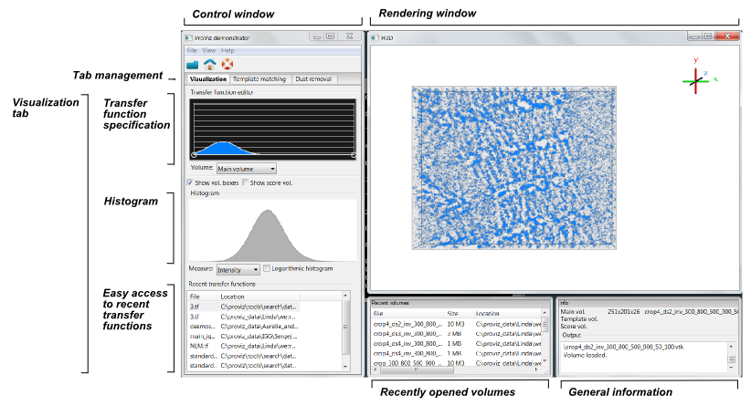
|
|
|
We are aiming to create a tool for visualizing nanometer resolution images of macromolecular structures such as proteins. It will work in the area between protein structural information, obtained from X-ray crystallography, and cell morphology information, obtained from conventional microscopy. In the ProViz project we combine the image acquisition technique Molecular Electron Tomography (MET) with advanced image analysis and visualization tools developed at the Centre for Image Analysis (CBA) in Uppsala. We will also make use of SenseGraphics technology for hapto-visual environments.
By using MET, which can be seen as the molecular biology equivalent to computed tomography in medical imaging, it is possible to observe the 3D conformation of proteins in cells. MET enables better translation of results between the human biology and lab bench research in model systems during drug discovery and development. The information about where and how proteins interact in situ obtainable with ProViz will facilitate taking one important step towards truly tailoring pharmaceutical drugs on a protein level. It will also give the research on structural biology the possibility to take a significant step further.
The purpose of ProViz is to observe the 3D conformation of proteins in cells and give the possibility for the user to see and feel the proteins in context in 3D and thereby obtaining a deeper knowledge of their complex, flexible structure.
Below is a screenshot from the current development version of the demonstrator. It includes different ways of interactively altering the visualization transfer function, to enhance features in the tomograms.

If you have been invited to test the demonstrator, you can download the latest version here:
Ongoing project started in 2008.
The Visualization Program by Knowledge Foundation, Vårdal Foundation, Foundation for Strategic Research, VINNOVA, and Invest in Sweden Agency.