Automated Image Acquisition and Analysis in the MiniTEM Instrument
Ida-Maria Sintorn, Gunilla Borgefors, Ewert Bengtsson
Partners: Vironova AB; Delong Instruments, Brno, Czech Republic
Funding: Eurostars project
Period: 1107-
Abstract: Transmission electron microscopy (TEM) is an important clinical diagnostic and material analysis tool. Transmission electron microscopes are expensive, complex, sensitive and bulky machines, often housed in specially built rooms to avoid vibrations affecting the imaging process.
Jointly with the partners Vironova and Delong Instruments the MiniTEM instrument has been developed in a Eurostars funded project. The MiniTEM instrument is a desk-top low voltage TEM designed for biological samples, with a high degree of automation regarding instrument alignment, image acquisition and analysis. It is a small, cheap, robust, and easy to use system that requires no more training than any simple lab equipment, and can be hosted in any office or lab (even mobile). Since the prototype launch in 2014, the instrument and the image acquisition and analysis methods have been further developed and fine-tuned.
During 2015, applications using automated image acquisition and subsequent analysis were presented at SCANDEM2015 - the Annual Conference of the Nordic Microscopy Society, Jyväskylä, Finland, and at MC 2015 - the Microscopy Conference, Göttingen, Germany. The MiniTEM instrument was also used for the TEM analysis in the paper "Asymmetric supercapacitors based on carbon nanofibre and polypyrrole/nanocellulose composite electrodes", published in RSC Advances.
Automated Multiscale Analysis of TEM Images for Improved Cost-Effective Diagnosis of Cilia Disorders
Amit Suveer, Nataša Sladoje, Joakim Lindblad, Ida-Maria Sintorn
Partners: Anca Dragomir, Anders Ahlander, Dept. of Immunology, Genetics and Pathology, UU
Funding: Biomed SPARC Project initiative
Period: 1504-
Abstract: Dysfunctional immotile cilia are often due to genetic disorders, and results in respiratory infections, reduced female fertility and infertility in males. Transmission Electron Microscopy (TEM) is the standard technique for diagnosing cilia disorders. A highly manual and time consuming procedure is performed by an expert pathologist at the microscope to set a diagnosis. In this project, we are aiming at developing approach for automating the TEM imaging and analysis to significantly reduce the time and efforts required by an expert pathologist for diagnosis and enables improved and more accurate diagnosis.
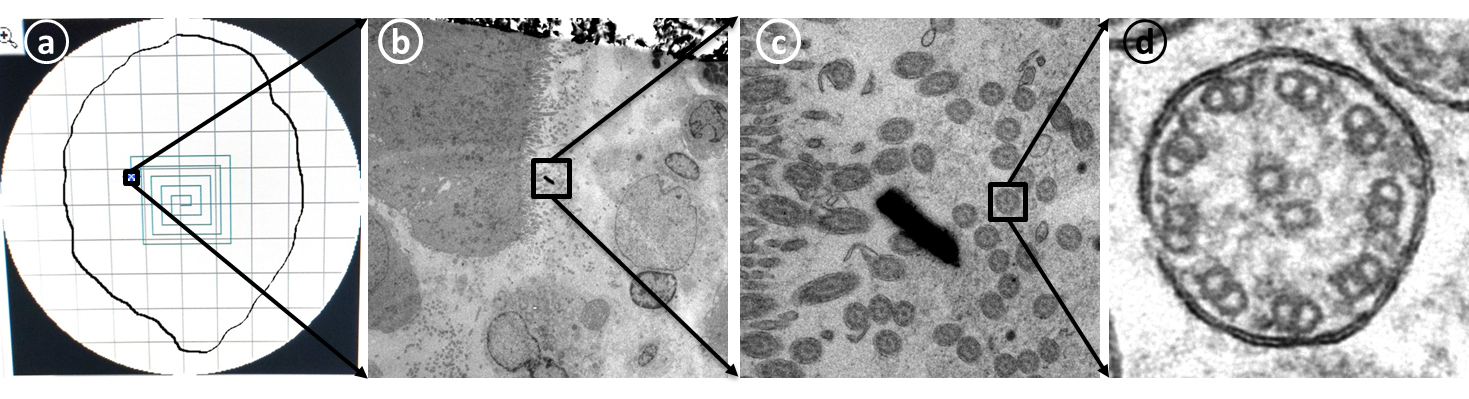
Our strategy includes automated navigation of a microscope for systematically traversing a specimen (illustrated in Fig. 25a), and acquire low magnification (LM) images, exemplified in Fig. 25b. A fast automated analysis of such low resolution images leads to detection of regions highly populated with cilia; one such region is shown in Fig. 25c (in a mid-magnification (MM), only used for illustration). Once such a region is detected, high magnification (HM) images, see Fig. 25d are acquired and used for the final cilia detection and super resolution reconstruction to enhance the fine structural details used to set a diagnosis.
The overview of the project, with some preliminary results was presented at Medicinteknikdagarna'15, Uppsala, Sweden in October 2015. Initial results related to the super resolution reconstruction were presented at the 5th International Conference on Image Processing Theory, Tools and Applications IPTA'15, Orleans, France in November 2015. Results related to analysis and cilia detection at LM will be presented at the International Symposium for Biomedical Imaging, ISBI'16, Prague, Czech Republic in April 2016.
|
CerviScan
Ewert Bengtsson, Bo Nordin
Partners: Rajesh Kumar, Centre for Development of Advanced Computing (CDAC), Thiruvananthapuram, Kerala, India; K. Sujathan, Regional Cancer Centre, Thiruvananthapuram, Kerala, India
Funding: Swedish Governmental Agency for Innovation Systems (VINNOVA); Swedish Research Council; SIDA
Period: 0801-
Abstract: Cervical cancer is a disease that annually kills over a quarter of a million women world-wide. This number could be substantially reduced if women were regularly screened for signs of cancer precursors using the well-established Pap-test. If detected early, these precursors can be treated with a very high rate of success. A problem with the Pap-test is that it requires highly trained cytotechnologists to perform the time consuming visual analysis of the specimen. For over 50 years attempts to automate this process have been made but still no cost effective systems are available.
The CerviScan project is an initiative from the Indian government, managed by the research institute CDAC in cooperation with the Regional Cancer Centre (RCC) in Kerala and CBA in Sweden, aimed at creating a low cost, automated screening system. The system will reduce the number of cytotechnologists needed for population screening by identifying and removing specimen that are clearly normal. A prototype system has been created and used to screen over 1000 specimen. Initial classification results are promising but screening times are still about 10 times longer than what is realistic in a real screening setting.
The original research funding has expired. Plans for the next phase of the project, focusing on dedicated hardware, are awaiting the result of funding applications in India and Sweden. In the meantime we have had funding for our collaboration from the Swedish Research Links Programme. The initial term for that funding expired at the end of 2015 but a new period has been granted for 2016-2018.
The project has resulted in several recent publications. Patrik Malm defended his PhD thesis closely linked to this project in February 2014. During 2015 two workshops were held within the project, one in India in March and one in Uppsala in October.
Analysis of Sperm Vitality
Ewert Bengtsson, Patrik Malm
Partners: Garcìa-Olalla, O., Alegre, E., Fernández-Robles, L. University of León, Industrial and Informatics Engineering School, León, Spain.
Funding: UU Faculty
Period: 13-15
Abstract: In our work on cervical cell image analysis in the CerviScan project we developed methods for Fourier shape analysis of cell nuclei. In collaboration with our partners in this project we tested whether those methods also could contribute to determine whether sperms to be used for artificial insemination were vital i.e. could be classified as acrosome-intact or acrosome-damaged. It turned out that the shape analysis when combined with previously used texture based features could lead to a classification accuracy of 99.13